
Written by Microscope Hunt Authority
Explore the hidden creatures around you with a microscope! Watch tiny creatures come to life, learn about science, and take stunning close-up photos
.
Introduction
Microscopes have for long been the link between the world we see with our eyes and the one that the naked human eye is unable to perceive. We are able to see objects and microorganisms that are not visible to the naked eye. Using this invention, scientists and students made amazing discoveries with the help of the microscope and its capabilities. From cells and bacteria to tiny mineral fragments and textile fibers, microscopes reveal details that deepen our understanding of the natural world. In this article, the microscope experiments that are suitable for students, beginners, and enthusiasts and which we will thoroughly describe will be the subject of our study. Each experiment will use simple, easy-to-find materials and will explain the observations in the easiest way possible yet with a professional tone.
Types of Microscopes Used in Experiments
Before diving into the experiments, it’s important to understand the different types of microscopes:
- Compound Microscope:
This is the most common microscope in classrooms. It uses two sets of lenses (objective and eyepiece) to magnify objects from 40x to 1000x. It is perfect for viewing cells, bacteria, and thin slices of tissues. - Stereo Microscope (Dissecting Microscope):
This microscope is used for observing larger, solid objects such as insects, leaves, and coins. It provides 3D views with lower magnification (10x to 40x). - Digital Microscope:
These microscopes connect to a computer or screen and often allow photo or video capture. They can be compound or stereo in design and are increasingly used in modern classrooms and homes.
Safety and Preparation
Before conducting any experiment, ensure you follow these safety tips:
- Always handle glass slides and cover slips carefully.
- Use gloves when dealing with biological materials.
- Clean your microscope lenses gently with lens paper.
- Start with the lowest magnification and adjust focus slowly.
- Work in a clean, well-lit environment.
Now, let’s begin the experiments!
Experiment 1: Observing Onion Cells
Materials:
- Onion
- Knife
- Tweezers
- Slide and cover slip
- Water
- Iodine solution (optional)
Procedure:
- Cut a small piece of onion and peel off a very thin, transparent layer of skin from the inner side.
- Place it on a slide and add a drop of water (or iodine if available).
- Gently place a cover slip on top.
- Observe under a compound microscope, starting with low magnification.
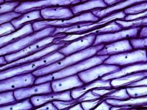
Observations:
You will see neatly arranged rectangular cells. The iodine helps stain the nucleus, making it more visible. Each cell has a visible cell wall and central nucleus.
Conclusion:
This classic experiment helps understand basic plant cell structure.
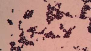
Experiment 2: Pond Water Life
Materials:
- Container of pond water
- Dropper
- Slides and cover slips
- Compound microscope
Procedure:
- Collect a small sample of pond water.
- Using a dropper, place a drop of water on the slide.
- Cover it with a cover slip.
- View under various magnifications.
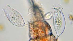
Observations:
Tiny organisms like protozoa, algae, or rotifers might be visible, moving actively. You might also see debris, plant fragments, or air bubbles.
Conclusion:
Pond water is teeming with microscopic life, demonstrating the diversity of unicellular organisms.
Experiment 3: Human Hair
Materials:
- A strand of human hair
- Slide and cover slip
- Water (optional)
- Stereo or compound microscope
Procedure:
- Place a strand of hair on the slide.
- Add a drop of water if needed, then cover with a slip.
- Observe under the microscope.
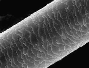
Observations:
You will notice the outer layer (cuticle), which may appear like overlapping scales. The thickness, color, and condition of hair become clear.
Conclusion:
This experiment shows the structural variation in hair and can be compared across individuals or animals.
Experiment 4: Cheek Cells
Materials:
- Clean cotton swab
- Slide and cover slip
- Methylene blue or iodine stain
- Water
- Compound microscope
Procedure:
- Gently rub the inside of your cheek with a cotton swab.
- Smear it on a slide.
- Add a drop of stain and water.
- Cover and observe.
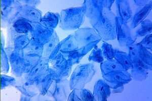
Observations:
Cheek cells appear round or irregular. The stained nucleus will be visible. The cell membrane and cytoplasm can also be seen.
Conclusion:
A great way to view human cells and understand cell structure.
Experiment 5: Mold on Bread
Materials:
- Moldy bread sample
- Slide and cover slip
- Tweezers
- Water
- Compound microscope
Procedure:
- Use tweezers to collect a small bit of mold.
- Place it on a slide, add a drop of water.
- Cover and observe.
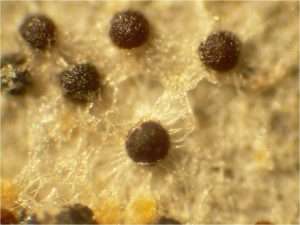
Observations:
Fungal hyphae (thread-like structures) and spore heads can be seen. The structures may appear fuzzy or branched.
Conclusion:
This experiment demonstrates fungal growth and reproduction.
Experiment 6: Salt and Sugar Crystals
Materials:
- Table salt
- Sugar
- Slides and cover slips
- Water (optional)
- Stereo or compound microscope
Procedure:
- Place a few grains of salt and sugar on separate slides.
- Add a drop of water to dissolve slightly if needed.
- Cover and observe.
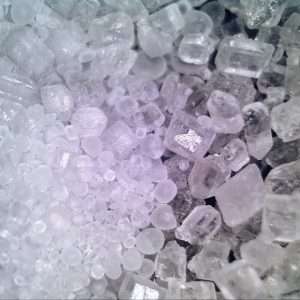
Observations:
Salt crystals usually appear cubic, while sugar crystals are more irregular. As they dissolve, edges become smoother.
Conclusion:
This highlights crystal structure and can introduce concepts of molecular arrangement.
Experiment 7: Fabric Fibers
Materials:
- Small fabric samples (cotton, wool, polyester)
- Scissors
- Slides or tape
- Stereo microscope
Procedure:
- Cut small threads of fabric.
- Mount them on a slide or stick them on tape.
- Observe under a stereo microscope.
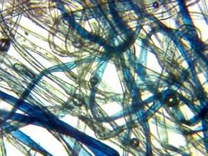
Observations:
You’ll notice differences in weave patterns, fiber smoothness, and thickness. Natural fibers usually look irregular, while synthetic fibers are uniform.
Conclusion:
Helps in understanding textile differences and microscopic material analysis.
Experiment 8: Leaf Structures and Stomata

Materials:
- Fresh leaf
- Nail polish (clear)
- Tape
- Slide
- Compound microscope
Procedure:
- Apply clear nail polish to the underside of the leaf.
- Let it dry completely.
- Press tape onto the dry polish and peel it off.
- Stick the tape to a slide and observe.
Observations:
You will see stomata (tiny pores) and epidermal cells. Stomata may appear as bean-shaped openings.
Conclusion:
This experiment demonstrates how plants breathe and exchange gases.
Experiment 9: Yeast in Sugar Water
Materials:
- Yeast
- Sugar
- Warm water
- Glass container
- Slides and cover slips
- Compound microscope
Procedure:
- Mix yeast, sugar, and warm water in a container.
- Wait for 10–15 minutes.
- Take a drop of the mixture and place it on a slide.
- Cover and observe.
Observations:
You may see yeast cells multiplying (budding). Some may be grouped or actively growing.
Conclusion:
Shows microbial growth and fermentation processes.
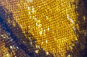
Experiment 10: Butterfly or Insect Wings
Materials:
- Butterfly/insect wing (found, not harmed)
- Slide and cover slip
- Stereo microscope
Procedure:
- Place a wing fragment on a slide.
- Cover gently if needed.
- Observe.
Observations:
Scales will appear like overlapping shingles. Colors may shimmer due to microscopic structure, not pigment.
Conclusion:
This reveals how insects use microscopic structures for color and insulation.
Care and Maintenance of the Microscope
To keep your microscope in good working condition:
- Store in a dust-free cover.
- Always start and end with the lowest magnification.
- Clean lenses with proper lens paper only.
- Do not use harsh chemicals.
- Handle slides carefully.
Conclusion
Experiments with microscopes are an experimental component to study biology chemistry and materials science. Microscope experiments offer an exciting and educational way to explore the world around us. Whether you’re looking at everyday objects or investigating the tiny life forms in pond water, microscope experiments can open your eyes to a whole new universe. These easy and fun experiments allow anyone to discover the wonders of the microscopic world with just a microscope and some simple materials. Dive into microscope experiments and start your journey into the unseen—there’s no limit to what you can discover!